SIMNRA VERSION 7.0
SIMNRA 7.0 offers the following improvements, compared to version 6.0:
- The program SigmaCalc 2.0 by A. Gurbich can be included, allowing calculating cross-section data for non-Rutherford scattering and nuclear reactions for many ion-target combinations at any angle.
- The program MottCalc by M. Kokkoris is included allowing to calculate the quantum-mechanical Mott cross-section for identical particles (requires SIMNRA version 7.03 or higher).
- Calculation of gamma-ray yields by particle-induced gamma-ray emission (PIGE) (requires SIMNRA version 7.02 or higher).
- Calculation of ERDA and NRA with incident neutrons (n-ERDA, n-NRA) (requires SIMNRA version 7.03 or higher).
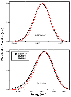
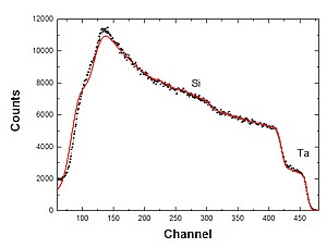
- Skewness of energy spread distributions: All energy spread distributions are approximated by two-piece normal distributions instead of Gaussian functions. This improves the accuracy of spectrum simulation by taking mean value, variance and third moment into account. See Fig. 1 for an example: The measured energy distributions are better approximated by the SIMNRA 7 model.
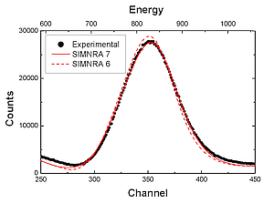
- Improved treatment of cross-sections with structure, see Fig. 2, resulting in improved accuracy for the simulation of non-Rutherford scattering and nuclear reactions.
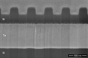
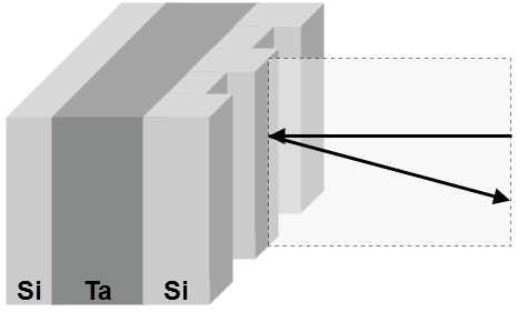
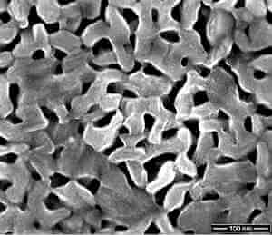
- Generalized layer roughness allows to describe various layer thickness distributions, see Fig. 3 for an example of a grating structure.
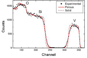
- Porous materials and layers, see Fig. 4.
- Arbitrary window for external beam.
- Energy-dependent detector sensitivity.
- Universal scattering cross-section based on the universal potential for simulation calculations at very low energies, for example for medium energy ion scattering (MEIS).
- Improved list of reactions with clearly arranged list of possible data.
- Possibility to resize experimental spectra to N channels; Optimized smoothing of experimental data.
- Modernized user interface with support for high-resolution devices until 200 dpi and support for multi-monitor systems.
- 32- and 64-bit versions of SIMNRA.
- Full Unicode support.
- Improved programming support: Full access to all properties of all SIMNRA objects through OLE automation.
- SIMNRA 7 uses the XML-based IBA data format as its native file format for storing spectra and all input data allowing easy exchange of data between different ion beam analysis programs.
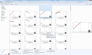
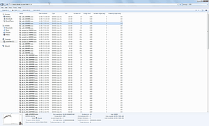
- Smooth integration into the Microsoft Windows environment by thumbnail providers and property handlers allowing to display thumbnail images of SIMNRA data files and providing additional information about the file content, see Fig. 5.
- Integration into Windows Search allows to search for specific files based on file content (for example ion species, beam energy), see Fig. 6.
A full list of changes from version 6.0 to version 7.0 can be found in the file CHANGES 7.0.TXT.
Download SIMNRA 7.0.